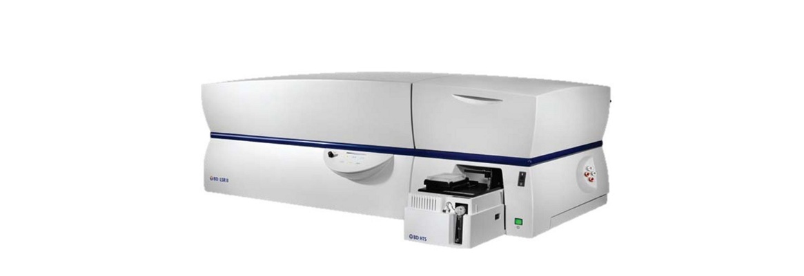
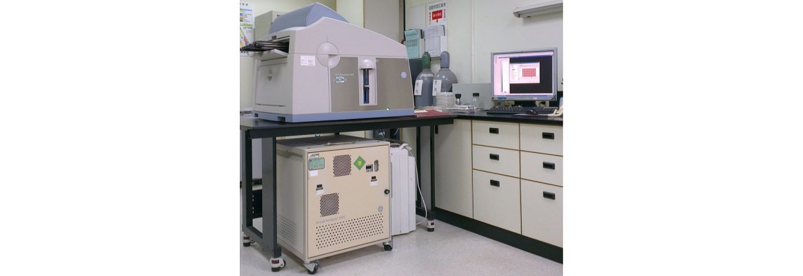
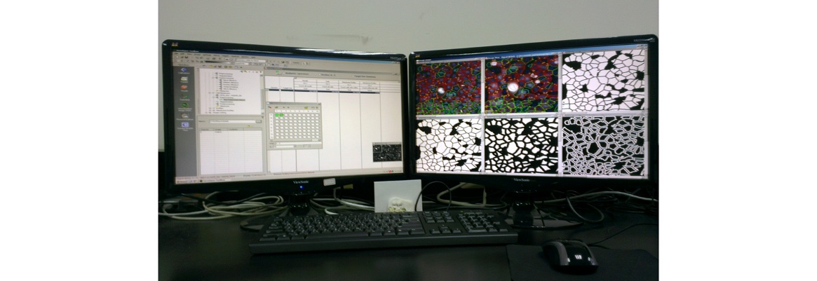
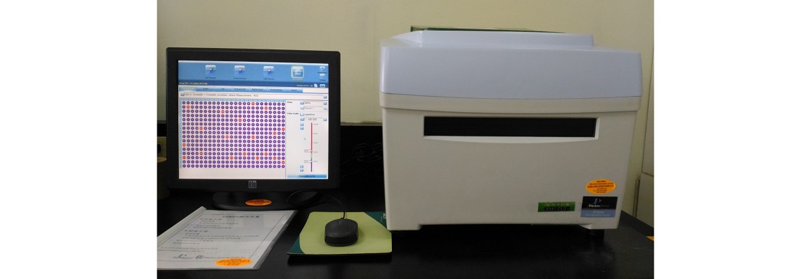
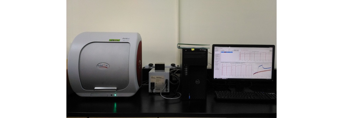
BD LSR II Cell Analyzer
BD LSR II Cell Analyzer
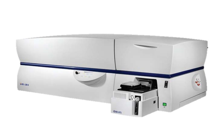
3-laser, 11 color (6-3-2)
|
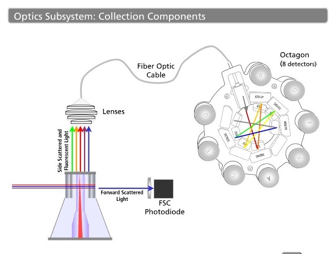
BD LSR II Configuration 3-laser, 11 color |
||
|
Excitation Laser |
Detection Filter |
example |
|
488nm Blue Laser |
530/30 |
Alexa Fluor 488, FITC, BB515 |
|
575/26 |
PE |
|
|
610/20 |
PE-Texas Red |
|
|
660/20 |
PE-Cy5 |
|
|
695/40 |
PI, PerCP, PerCP-Cy5.5, BB700, 7-AAD |
|
|
780/60 |
PE-Cy7 |
|
|
633nm Red Laser |
670/30 |
APC, Alexa Fluor 647 |
|
730/45 |
Alexa Fluor 700 |
|
|
780/60 |
APC-Cy7, APC-H7 |
|
|
405nm Violet Laser |
450/50 |
Alexa Fluor 405, V450, BV421, PB |
|
525/50 |
Alexa Fluor 430, V500, BV510 |
|
Consumables
(1) 5-mL polystyrene test tubes, 12 x 75-mm (BD Falcon Cat. No.352052)
1. Sample preparation
(1) Recommend sample concentration: 1x10⁶ cells /ml
(2) Filter your samples
Recommend filtering samples through a nylon mesh 40 µm (BD Cat. No. 352340) or 5-mL polystyrene test tubes, 12 x 75-mm (BD Falcon) with cell-strainer cap (BD Cat. No. 352235) if aggregated cells can be seen in the sample tube.
Filtered sample should be saved on ice, protected from light.
Data
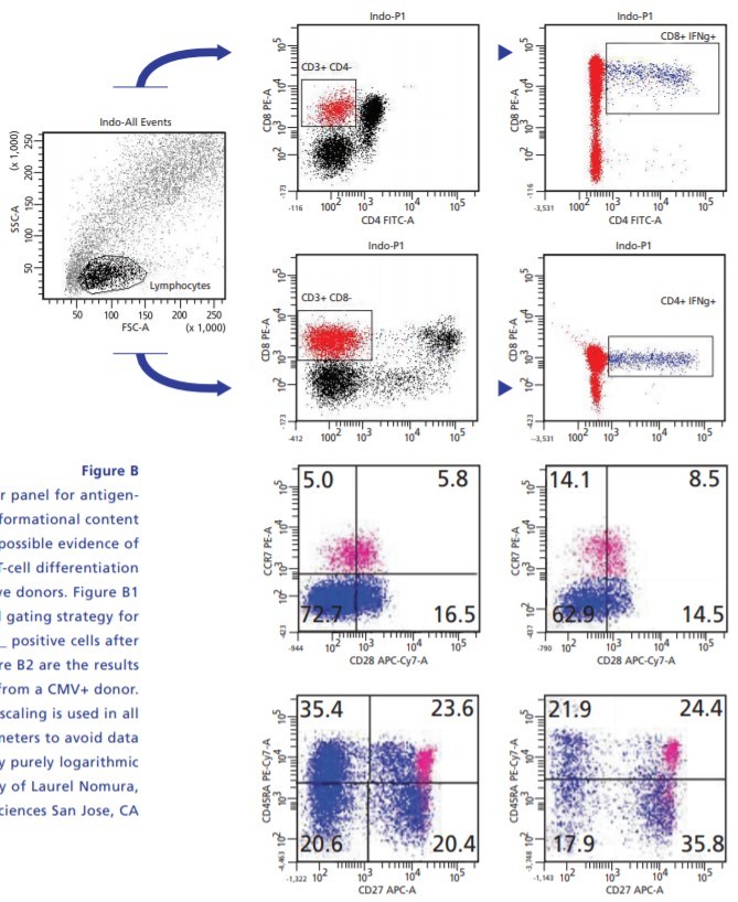
8-color panel for Antigenspecific Phenotyping
Using an eight-color panel for antigenspecific phenotyping, informational content is maximized providing possible evidence of novel patterns of T-cell differentiation among CMV-responsive donors. Figure B1 shows a sequential gating strategy for phenotyping of IFN_ positive cells after CMV simulations. Figure B2 are the results of total CD8 T cells from a CMV+ donor.
|
|
|
|
Immunophenotyping
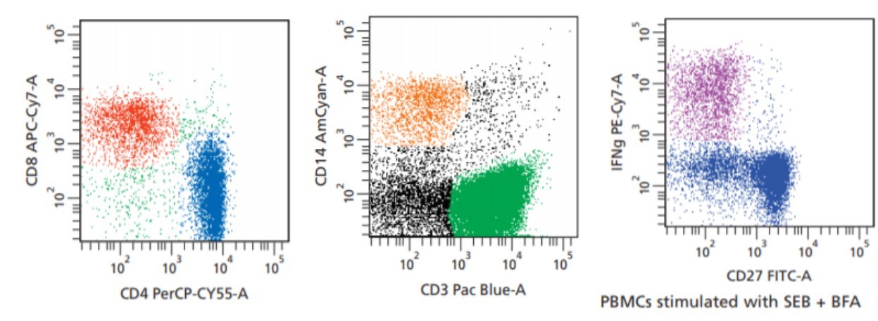
Characterizing cells at different stages of development through the use of fluorescent-labeled monoclonal antibodies against surface markers is one of the most common applications of flow cytometry. Surface staining and intracellular staining are common application for flow cytometry. PBMCs stimulated with SEB and protein transport inhibitor -BFA and detect the expression of IFN-γ.
Attachment:
2. FACSDivav6_LSRII_quickguide
Reference website: BD Bioscience
https://www.bd.com/resource.aspx?IDX=17868
http://www.indiana.edu/~fccf/pdf/LSRIIBrochure.pdf
